Saturday, November 17, 2018

ECG or EKG Basics

You have access to ECG's if you have a surgical monitor with 3 clips that are put on as shown here:

These clips are pretty cool.  Even though they are clipped to the skin high up on the pet's legs, they pick up the electrical impulses generated by the heart as it beats and conducts them through wires to the monitor which displays them as a wavy line that should look something like this:


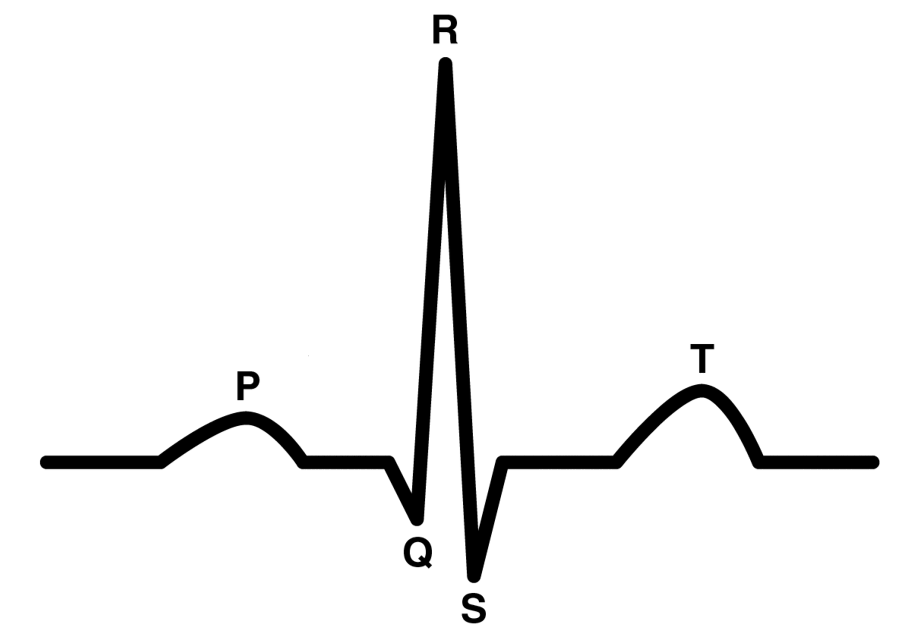
So, what do those waves mean?  Well, let's label them so we can discuss each part of the wave individually:
Okay, now we can start finally start talking about the heart!


The heart beats like this:
1.  the atria contract (these are the small chambers on the "top" of the heart)
2.  the ventricles contract (these are the larger chambers on the "bottom" of the heart)

The blood flows like this:
1. de-oxygenated blood enters the right atrium
2. when the atrium contracts the blood is pumped into the right ventricle
3. when the right ventricle contracts the blood is pumped into the lungs and oxygenated there
4. the blood comes back from the lungs and enters the left atrium
5. when the left atrium contracts the blood is pumped into the left ventricle
6. when the left ventricle contracts, the oxygenated blood is pumped out to the whole body

The electrical activity of the heart flows like this:
1. the SA node is the "pacemaker of the heart" and is located in the right atrium, it fires and the atria both contract, this is the "P" wave as shown above.
2. the electrical impulse then travels through the center of the heart and down to the ventricles, when they contract you see the "Q","R", and "S" waves shown above.
3. the electrical impulse then re-polarizes or "resets" which creates the "T" wave shown above.

Interpreting abnormal ECG's:


Above we see a normal beat (a P, Q, R, S, and T), another normal wave, and then a long pause followed by a Q, R, S, T and then another normal beat.  So, that 3rd set is preceded by a long pause and is also missing it's "P" wave.  What does this mean?

The "P" is where the electrical activity starts.  The SA node fires and the atria attract.  There is an issue here.  This didn't happen during this beat.  When the ventricles don't get a signal to contract they wait for a little bit and then, rather than not beating at all, they'll fire on their own.  So, the impulse to beat didn't happen so the ventricles fired on their own.  This is called an "escape beat".


Above we see a T wave (the end of the previous beat), then a normal P, Q, R, S, T followed by a P-wave without a Q, R, S, and T.  Then another P-wave all alone, then the next complex is normal (P, Q, R, S, and T).  After that is another P-wave without a Q, R, S, and T, then 2 more normal complexes.  What is happening here?

Well, when there is a P-wave that is not followed by a Q, R, and S you have an electrical signal that is firing the atria, but then not getting passed along to the ventricles to cause them to contract.  This is called "heart block" -an appropriate name since the signal is getting blocked between the atria and ventricles somewhere in the middle of the heart!

The above is a very brief although only slightly simplified introduction to ECG's.  For more information I highly recommend checking out: http://ekg.academy/ where they have lessons and lots of practice leads to nerd out on!

Anesthesia -Oxygen Flow Rate

There is a lot of disagreement out there on how to set the oxygen flow rate when running anesthesia.  Individual clinics and techs seem to have their own rule of thumb, but here's a review of how the anesthesia machine works, how to monitor ETCO2 and how to determine if you're wasting oxygen, your anesthetic agent (such as isoflurane), and potentially adding to your patients' hypothermia.

Assuming a rebreathing system, no ventilator, and just an oxygen tank (no nitrogen, etc):

When your patient is hooked up the only gas available to him or her is oxygen.  That oxygen has 2 functions, for profusion of tissues (to keep the pet alive) and to carry your anesthetic gas.

How do you know if your O2 flow rate is too low?

There is no other gas in the system other than oxygen and the exhaled CO2 of your patient.  So, with O2 flow rates that are too low you'd see:
  • difficulty stabilizing anesthesia level because below 0.5L/min most vaporizors cannot reliably deliver anesthetic gas at the set percentage
  • the reservoir bag would not be inflated
  • the inhaled or IN CO2 would be above 0
  • ETCO2 would steadily increase as CO2 builds up in the circle faster than the O2 can replace it
  • SpO2 would begin to decrease
  • mucous membrane color would turn cyanotic
  • patient death

How do you know if your O2 flow rate is too high?

  • you're going through oxygen tanks too quickly
  • your reservoir bag is fully inflated
  • you're going through your anesthetic gas too quickly
  • your patient gets cold despite your warming efforts
Clearly a balance is needed, and you want to error on the side of too high of a flow rate.  Most clinics use a default O2 flow rate of 1L/min for anything under 100lbs and that works well.  Others calculate the rate based on 30ml/kg with a minimum of 500ml/min based on their vaporizor's limit for minimum O2 flow.

Common myths:

  • Increasing O2 flow rate will decrease ETCO2.
    • If the patient is not inhaling any CO2 than the cause of a high ETCO2 is inadequate ventilation.  This means the rate or quality of breaths is the issue.  Increasing the amount of oxygen available won't change ETCO2 in this case.  Manual ventilation should be used to decrease the ETCO2 and anesthesia depth should be evaluated to see if the cause of inadequate ventilation is excessive anesthesia depth.
  • Increasing the O2 flow rate (alone) will change anesthesia depth.
    • While it is true that turning the oxygen flow rate up or down will also effect the amount of anesthetic gas in the system, the amount of anesthetic gas breathed in by the patient will remain the same unless you also change your vaporizer setting.  This is because the amount of gas present is a percentage of the oxygen being delivered so it is flowing out of the system faster or slower too.  It's like turning a hose on in a box with a hole in it designed to keep the box 2% full at all times, then turning the hose up and having the hole get bigger at the same time -you're just using more water, but the box stays 2% full.
    • NOTE:  It is true that increasing the oxygen flow rate for a short period of time while making changes in the anesthetic gas percentage will help the change take place faster since it will push the gas out of the system faster.

Cautions:


  • If your patient is not adequately ventilating and you start giving him manual breaths, along with an increase of oxygen he/she will also be getting more anesthetic gas (because your oxygen is carrying a certain percentage of anesthetic gas, so more O2 means more gas).  If the reason for inadequate ventilation was excessive depth, manually breathing for the patient could make the problem worse, so be sure to turn the vaporizer down and flush the system prior to PPV.

Getting your dog to accept having his teeth brushed

Obviously not all dogs will allow their owners to brush their teeth, but it is my firm belief that more dogs will either accept it (or love ...